Negative staining
In principle, the specimen is surrounded by heavy metal atoms that act as an electron barrier. Because the electron beam can pass through the low electron density of the specimen but not through the metallic background, the result is negative staining: a light specimen against dark background.
pH and concentration of the staining solution and the duration of staining varies according to specimen types. The optimal conditions for staining are found by trial and error. Routinely used negative staining solutions are
- Phosphotungstic acid (KPT), 1% pH 6.5 and 2% pH 7.5
- 1% Uranyl acetate in water
- (also available 3% UA in water, or 2% neutralized uranyl acetate)
- 5% Ammonium mobybdate - 1% trehalose, pH 7.4
Medium of astrocytes was collected and concentrated, and the peak fractions containing cholesterol were subjected to negative staining. The size distribution of the particles is shown. Bar is 100 nm. (Mutka et al., 2004, J.Biol.Chem. 279:48654-48662).
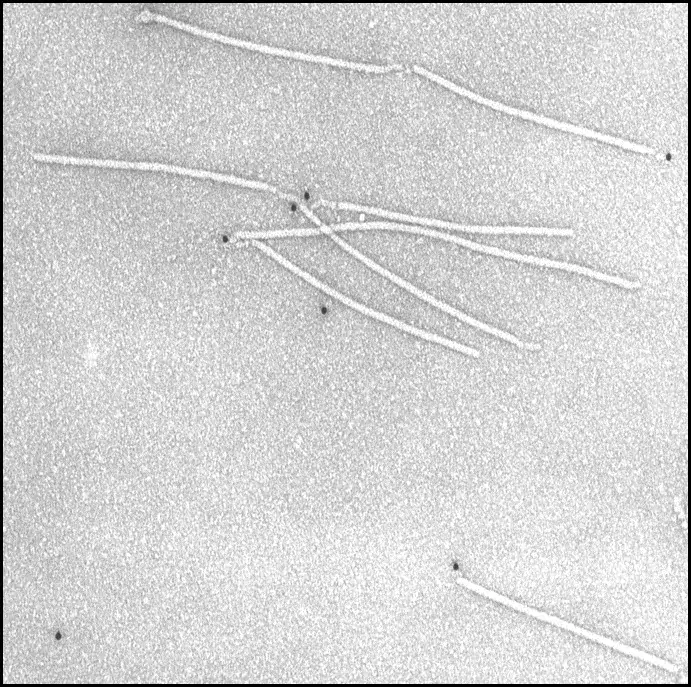
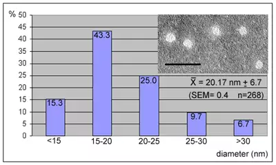
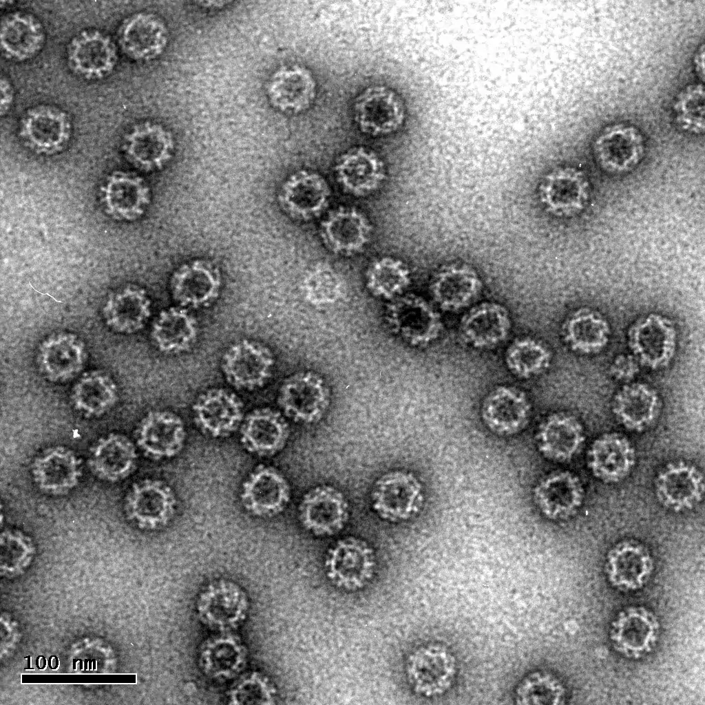
Image 1 Detection of Potyviral genome-linked protein VPg at only one end of the virions (Puustinen et al., 2002, J.Virol. 76:12703-12711)
Image 2 Whole mount immunolabelling is a method that combines immunolabelling and negative staining. Specimen is applied to the grid and immunolabelled prior negative staining.