Correlative Light Electron Microscopy
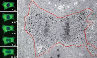
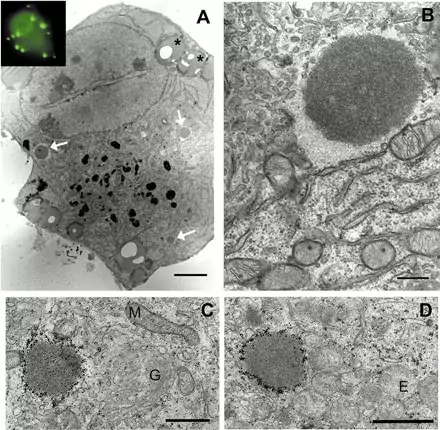
Image 1 In fluorescence images (left) a dividing cell is progressing from metaphase to anaphase, and then the cell was fixed and processed for immunoEM. Green colour and black precipitate represent GFP-tagged ER-protein and blue colour chromosomes. (M. Puhka).
Image 2 Epifluorescence image (inset in A) and low power TEM image (A) of the same primary sympathetic neuron showing that over-expressed GFP-XIAP is accumulated into the inclusion bodies (bright spots in inset, arrows in A and high-power image of one of the structures in B). Immunolabelling using anti-GFP antibodies verified that the inclusion bodies found in TEM images corresponded to the bright spots imaged with fluorescence microscope (Yu et al., 2003, MCN 22:308-318).